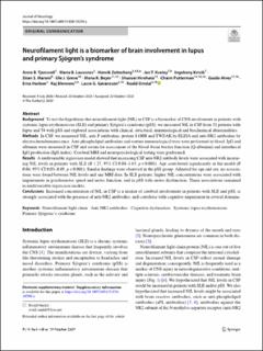
Neurofilament light is a biomarker of brain involvement in lupus and primary Sjögren’s syndrome
| dc.contributor.author | Tjensvoll, Anne Bolette | |
| dc.contributor.author | Lauvsnes, Maria Boge | |
| dc.contributor.author | Zetterberg, Henrik | |
| dc.contributor.author | Kvaløy, Jan Terje | |
| dc.contributor.author | Kvivik, Ingeborg | |
| dc.contributor.author | Maroni, Stian | |
| dc.contributor.author | Greve, Ole Jacob | |
| dc.contributor.author | Beyer, Mona K. | |
| dc.contributor.author | Hirohata, Shunsei | |
| dc.contributor.author | Putterman, Chaim | |
| dc.contributor.author | Alves, Guido Werner | |
| dc.contributor.author | Harboe, Erna | |
| dc.contributor.author | Blennow, Kaj | |
| dc.contributor.author | Gøransson, Lasse | |
| dc.contributor.author | Omdal, Roald | |
| dc.date.accessioned | 2020-11-20T11:24:23Z | |
| dc.date.available | 2020-11-20T11:24:23Z | |
| dc.date.created | 2020-11-19T16:21:39Z | |
| dc.date.issued | 2020-10 | |
| dc.identifier.citation | Tjensvoll, A.B, Lavusnes, M.B., Zetterberg, H. (2020) Neurofilament light is a biomarker of brain involvement in lupus and primary Sjögren’s syndrome. Journal of Neurology. https://doi.org/10.1007/s00415-020-10290-y | en_US |
| dc.identifier.issn | 0340-5354 | |
| dc.identifier.uri | https://hdl.handle.net/11250/2688884 | |
| dc.description.abstract | Background To test the hypothesis that neurofilament light (NfL) in CSF is a biomarker of CNS involvement in patients with systemic lupus erythematosus (SLE) and primary Sjögren’s syndrome (pSS), we measured NfL in CSF from 52 patients with lupus and 54 with pSS and explored associations with clinical, structural, immunological and biochemical abnormalities. Methods In CSF, we measured NfL, anti-P antibodies, protein S100B and TWEAK by ELISA and anti-NR2 antibodies by electrochemiluminescence. Anti-phospholipid antibodies and routine immunological tests were performed in blood. IgG and albumin were measured in CSF and serum for assessment of the blood–brain barrier function (Q-albumin) and intrathecal IgG production (IgG index). Cerebral MRI and neuropsychological testing were performed. Results A multivariable regression model showed that increasing CSF anti-NR2 antibody levels were associated with increasing NfL levels in patients with SLE (B 1.27, 95% CI 0.88–1.65, p < 0.001). Age contributed significantly in the model (B 0.04, 95% CI 0.03–0.05, p < 0.001). Similar findings were observed in the pSS group. Adjusted for age and sex, no associations were found between NfL levels and any MRI data. In SLE patients, higher NfL concentrations were associated with impairments in psychomotor speed and motor function, and in pSS with motor dysfunction. These associations remained in multivariable regression models. Conclusions Increased concentration of NfL in CSF is a marker of cerebral involvement in patients with SLE and pSS, is strongly associated with the presence of anti-NR2 antibodies, and correlates with cognitive impairment in several domains. | en_US |
| dc.language.iso | eng | en_US |
| dc.publisher | Springer | en_US |
| dc.rights | Navngivelse 4.0 Internasjonal | * |
| dc.rights.uri | http://creativecommons.org/licenses/by/4.0/deed.no | * |
| dc.subject | Sjögren’s syndrome | en_US |
| dc.subject | Lupus | en_US |
| dc.title | Neurofilament light is a biomarker of brain involvement in lupus and primary Sjögren’s syndrome | en_US |
| dc.type | Peer reviewed | en_US |
| dc.type | Journal article | en_US |
| dc.description.version | publishedVersion | en_US |
| dc.rights.holder | © The Author(s) 2020 | en_US |
| dc.subject.nsi | VDP::Medisinske Fag: 700::Klinisk medisinske fag: 750::Nevrologi: 752 | en_US |
| dc.source.pagenumber | 10 | en_US |
| dc.source.journal | Journal of Neurology | en_US |
| dc.identifier.doi | 10.1007/s00415-020-10290-y | |
| dc.identifier.cristin | 1850057 | |
| cristin.ispublished | true | |
| cristin.fulltext | original | |
| cristin.qualitycode | 2 |
Tilhørende fil(er)
Denne innførselen finnes i følgende samling(er)

Med mindre annet er angitt, så er denne innførselen lisensiert som Navngivelse 4.0 Internasjonal