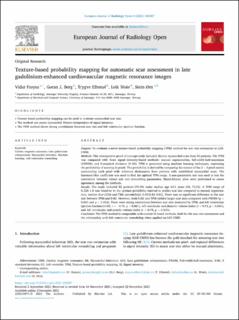
Texture-based probability mapping for automatic scar assessment in late gadolinium-enhanced cardiovascular magnetic resonance images
| dc.contributor.author | Frøysa, Vidar | |
| dc.contributor.author | Berg, Gøran Jansson | |
| dc.contributor.author | Eftestøl, Trygve Christian | |
| dc.contributor.author | Woie, Leik | |
| dc.contributor.author | Ørn, Stein | |
| dc.date.accessioned | 2021-12-21T10:23:49Z | |
| dc.date.available | 2021-12-21T10:23:49Z | |
| dc.date.created | 2021-12-10T19:26:39Z | |
| dc.date.issued | 2021-12 | |
| dc.identifier.citation | Frøysa, V., Berg, G.J., Eftestøl, T., Woie, L., Ørn, S. (2021) Texture-based probability mapping for automatic scar assessment in late gadolinium-enhanced cardiovascular magnetic resonance images. European Journal of Radiology Open, 8, 100387 | en_US |
| dc.identifier.issn | 2352-0477 | |
| dc.identifier.uri | https://hdl.handle.net/11250/2835215 | |
| dc.description.abstract | Purpose To evaluate a novel texture-based probability mapping (TPM) method for scar size estimation in LGE-CMRI. Methods This retrospective proof-of-concept study included chronic myocardial scars from 52 patients. The TPM was compared with three signal intensity-based methods: manual segmentation, full-width-half-maximum (FWHM), and 5-standard deviation (5-SD). TPM is generated using machine learning techniques, expressing the probability of scarring in pixels. The probability is derived by comparing the texture of the 3 × 3 pixel matrix surrounding each pixel with reference dictionaries from patients with established myocardial scars. The Sørensen-Dice coefficient was used to find the optimal TPM range. A non-parametric test was used to test the correlation between infarct size and remodeling parameters. Bland-Altman plots were performed to assess agreement among the methods. Results The study included 52 patients (76.9% male; median age 64.5 years (54, 72.5)). A TPM range of 0.328–1.0 was found to be the optimal probability interval to predict scar size compared to manual segmentation, median dice (25th and 75th percentiles)): 0.69(0.42–0.81). There was no significant difference in the scar size between TPM and 5-SD. However, both 5-SD and TPM yielded larger scar sizes compared with FWHM (p < 0.001 and p = 0.002). There were strong correlations between scar size measured by TPM, and left ventricular ejection fraction (LVEF, r = −0.76, p < 0.001), left ventricular end-diastolic volume index (r = 0.73, p < 0.001), and left ventricular end-systolic volume index (r = 0.75, p < 0.001). Conclusion The TPM method is comparable with current SI-based methods, both for the scar size assessment and the relationship with left ventricular remodeling when applied on LGE-CMRI. | en_US |
| dc.language.iso | eng | en_US |
| dc.publisher | Elsevier Ltd. | en_US |
| dc.relation.uri | http://dx.doi.org/10.1016/j.ejro.2021.100387 | |
| dc.rights | Attribution-NonCommercial-NoDerivatives 4.0 Internasjonal | * |
| dc.rights.uri | http://creativecommons.org/licenses/by-nc-nd/4.0/deed.no | * |
| dc.subject | maskinlæring | en_US |
| dc.subject | kardiologi | en_US |
| dc.title | Texture-based probability mapping for automatic scar assessment in late gadolinium-enhanced cardiovascular magnetic resonance images | en_US |
| dc.type | Peer reviewed | en_US |
| dc.type | Journal article | en_US |
| dc.description.version | publishedVersion | en_US |
| dc.rights.holder | © 2021 The Authors. | en_US |
| dc.subject.nsi | VDP::Teknologi: 500::Medisinsk teknologi: 620 | en_US |
| dc.subject.nsi | VDP::Medisinske Fag: 700::Klinisk medisinske fag: 750::Kardiologi: 771 | en_US |
| dc.source.volume | 8 | en_US |
| dc.source.journal | European Journal of Radiology Open (EJR Open) | en_US |
| dc.identifier.doi | 10.1016/j.ejro.2021.100387 | |
| dc.identifier.cristin | 1967284 | |
| dc.source.articlenumber | 100387 | en_US |
| cristin.ispublished | true | |
| cristin.fulltext | original | |
| cristin.qualitycode | 1 |
Files in this item
This item appears in the following Collection(s)

Except where otherwise noted, this item's license is described as Attribution-NonCommercial-NoDerivatives 4.0 Internasjonal