Defining angular and radial positions and parameters for myocardial pixels in cardiac MR images
Journal article, Peer reviewed
Åpne
Permanent lenke
http://hdl.handle.net/11250/2374856Utgivelsesdato
2014Metadata
Vis full innførselSamlinger
Originalversjon
Engan, K., Woie, L. & Eftestøl, T. (2014) Defining angular and radial positions and parameters for myocardial pixels in cardiac MR images. Computing in Cardiology, 41Sammendrag
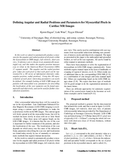
In this work we aimed to automatically produce a measure
for the angular and radial position of all pixels within
the myocardium in CMR images, left ventricle, short axis
view. A reference axis is chosen in an anatomically meaningful
way, and this makes the angular position measure
easy to relate to the American Heart Association (AHA)
17 segment model. The angular and the radial position
give values for each pixel so that each pixel can be represented
by a 3D vector of information [intensity value,
angular position, radial position]. Using this 3D representation
of each pixel, interesting parameters can easily
be defined. For example looking at LGE-CMR images for
patients with myocardial scar, parameters for describing
the localization of the scar segments can be found automatically
and objectively, and can be used for further classification
of patients.
Beskrivelse
The article was originally published under a creative commons license in 2014 in Computing in Cardiology; http://www.cinc.org/archives/2014/.
Utgiver
IEEETidsskrift
Computing in Cardiology
Med mindre annet er angitt, så er denne innførselen lisensiert som Navngivelse 3.0 Norge