Optical coherence tomography features and risk of macular hole formation in the fellow eye
| dc.contributor.author | Lindtjørn, Birger | |
| dc.contributor.author | Krohn, Jørgen Gitlesen | |
| dc.contributor.author | Forsaa, Vegard A. | |
| dc.date.accessioned | 2023-03-01T10:54:59Z | |
| dc.date.available | 2023-03-01T10:54:59Z | |
| dc.date.created | 2021-10-14T15:03:46Z | |
| dc.date.issued | 2021 | |
| dc.identifier.citation | Lindtjørn, B., Krohn, J., & Forsaa, V. A. (2021). Optical coherence tomography features and risk of macular hole formation in the fellow eye. BMC ophthalmology, 21(1), 1-7. | en_US |
| dc.identifier.issn | 1471-2415 | |
| dc.identifier.uri | https://hdl.handle.net/11250/3054911 | |
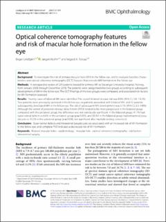
| dc.description.abstract | Background To investigate the risk of primary macular hole (MH) in the fellow eye, and to evaluate baseline characteristics and optical coherence tomography (OCT) features that precede MH formation in the fellow eye. Methods A retrospective review of 229 patients treated for primary MH at Stavanger University Hospital, Norway, from January 2008 through December 2018. The patients were categorised into two groups according to subsequent development of MH in the fellow eye. The OCT findings of the two groups were compared, and associated risk factors for MH formation assessed. Results Twenty cases of bilateral MH were identified. The overall bilateral disease risk was 8.8% (95% CI, 5.8–13.2%). Two patients were previously operated in the fellow eye, six patients presented with bilateral MH, and 12 patients subsequently developed MH in the fellow eye. The risk of subsequent MH development was 5.7% (95% CI, 3.3–9.8%). Although the extent of posterior vitreous detachment (PVD) tended to be more progressed in the bilateral group compared with the unilateral group, the difference was not statistically significant. In the bilateral group, 41.7% had outer retinal defects vs 6.6% in the unilateral group (p = 0.001), and 33.3% in the bilateral group had intraretinal pseudocysts vs 10.2% in the unilateral group (p = 0.036, not significant after multiple testing correction). Conclusion Outer retinal defects and intraretinal pseudocysts are associated with an increased risk of MH formation in the fellow eye, and complete PVD indicates a decreased risk of MH formation. | en_US |
| dc.language.iso | eng | en_US |
| dc.publisher | BMC | en_US |
| dc.rights | Navngivelse 4.0 Internasjonal | * |
| dc.rights.uri | http://creativecommons.org/licenses/by/4.0/deed.no | * |
| dc.title | Optical coherence tomography features and risk of macular hole formation in the fellow eye | en_US |
| dc.type | Peer reviewed | en_US |
| dc.type | Journal article | en_US |
| dc.description.version | publishedVersion | en_US |
| dc.rights.holder | The authors | en_US |
| dc.subject.nsi | VDP::Medisinske Fag: 700 | en_US |
| dc.source.pagenumber | 1-7 | en_US |
| dc.source.volume | 21 | en_US |
| dc.source.journal | BMC Ophthalmology | en_US |
| dc.source.issue | 1 | en_US |
| dc.identifier.doi | 10.1186/s12886-021-02111-1 | |
| dc.identifier.cristin | 1946031 | |
| cristin.ispublished | true | |
| cristin.fulltext | original | |
| cristin.qualitycode | 1 |
Tilhørende fil(er)
Denne innførselen finnes i følgende samling(er)

Med mindre annet er angitt, så er denne innførselen lisensiert som Navngivelse 4.0 Internasjonal